Excessive epithelial mechanosensation drives nociceptive innervation and chronic bladder pain via the PIEZO1-SLC7A11-glutamate axis
个人文献精读 Research Note,用于梳理论文证据链、图像逻辑、补充材料与可迁移研究思路。
Min Jung Kim; Joo Hwan Noh; Ho Young Lee; Bohm Lee; Alexander Doh Park; Hyeonseo Oh; Geun-Woo D. Kim; Hye-Sook Lee; Yeonsoo Oh; Hyeonje Hwang; Chungoo Kim; Hyeonjong Lee; Xue Li; Mi Mi Oh; Donghoon Lim; Soman N. Abraham; Yongcheol Cho; Sung Wook Chi; Hae Woong Choi
Cell 189, 1-22 (2026; corrected proof)
Open access under CC BY 4.0;© 2026 The Author(s), published by Elsevier Inc. 公开版按开放获取与导读引用边界处理。
Morgen · tomorgene.com
公开导读版:7 张低分辨率代表图 + 主图/补图占位索引;robots noindex,nofollow。
一句话总结
这篇文章把慢性膀胱感染后的疼痛问题重写成一条 neuroepithelial signaling chain:IL-6 将上皮 PIEZO1 调高,机械刺激被放大成 Ca2+/ROS,NRF2-SLC7A11 抗氧化反应释放 glutamate,最终驱动 nociceptive C-fiber sprouting、慢性骨盆痛样状态和排尿功能异常。
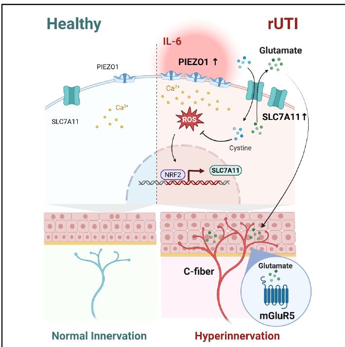
Graphical abstract:IL-6-PIEZO1-SLC7A11-glutamate axis 连接上皮机械应激与慢性膀胱疼痛。
名词解释
核心词支撑主图阅读;模型词帮助定位实验场景;方法词用于回到图注、methods 和 supplementary tables 检索。
核心机制 读懂主图必备
recurrent uropathogenic E. coli infection;本文用三次膀胱腔内感染模拟反复感染暴露和慢性疼痛样状态。
膀胱上皮感受充盈/牵张并向感觉神经传递信号;正常用于排尿反射,病理状态下变成疼痛放大器。
机械敏感 Ca2+ channel;IL-6/STAT3 上调后让 BEC 对 YODA1 或 stretch 更敏感。
感染炎症信号把免疫状态写入 PIEZO1 promoter,是机械敏感性被重新设定的 upstream priming。
PIEZO1 介导 Ca2+ influx 后增加 ROS;这是从机械刺激到 redox stress 的桥。
ROS 触发 NRF2 入核和抗氧化基因表达;在这里本来是保护反应,却引出疼痛链条。
cystine-glutamate antiporter,进口 cystine 维持抗氧化,出口 glutamate 改变神经上皮微环境。
上皮释放 glutamate 激活 DRG sensory neuron 的 metabotropic glutamate receptor 5,促进 neurite sprouting 和敏化。
substance P、CGRP、beta III-tubulin 标记的感觉纤维异常进入/靠近 urothelium,是慢性疼痛样状态的结构基础。
感染后的上皮屏障、机械通道、代谢分泌和感觉神经结构一起改变,而不是单一炎症指标上升。
实验模型/场景 看图时定位场景
WT female mice 每周 UPEC CI5 腔内感染三次;NU14/UTI89 作为 strain generalization。
人 BEC 体外系统,用于 MOI infection、IL-6/YODA1、PIEZO1/SLC7A11 CRISPR 和 glutamate release。
urothelium-specific Piezo1 cKO,用于证明上皮 PIEZO1 是 SLC7A11 induction、神经重塑和排尿异常的上游。
urothelium-specific Slc7a11 cKO,用于证明上皮 SLC7A11 是 glutamate/pain/voiding phenotype 的必要环节。
用来验证炎症 priming 是否必要;缺 IL-6 时 PIEZO1/SLC7A11/neuroplasticity/pain response 均被削弱。
胚胎 DRG 做 neurite outgrowth 和 Sholl analysis,成年 L6-S1 DRG 做 bladder-innervating sensory neuron marker。
sulfasalazine 与 sorafenib tosylate 作为 SLC7A11/glutamate release 抑制策略,测试疼痛和排尿功能 rescue。
awake bladder pressure 与 external urethral sphincter EMG 联合读出,连接疼痛反射和 voiding dysfunction。
方法与数据口径 遇读再查
用腹部机械刺激响应频率衡量 pelvic mechanical hypersensitivity;重点看 4 g 和 6 g response。
用尿斑数量、总面积、primary void/total area 衡量尿频/排尿组织性。
ex vivo transwell bladder mucosa barrier readout,帮助说明感染后上皮屏障性质改变。
BEC 或 bladder mucosal sheet 中的 Ca2+ influx 读出,用于 YODA1/stretch/感染状态比较。
general intracellular ROS readout;要和 Ca2+ influx、YODA1、PIEZO1 KO 一起解读。
验证 IL-6、PIEZO1、SLC7A11、p-STAT3、NRF2 等表达/激活。
PIEZO1 promoter STAT3 motif deletion 证明 IL-6/STAT3 对转录调控的直接性。
bulk infected bladder RNA-seq 与公开 cyclophosphamide injury scRNA-seq 联合定位 cycling/superficial urothelial response。
量化 eDRG neurite branching complexity,判断 glutamate/mGluR5 对结构塑性的作用。
S1-S3 是 sgRNA、shRNA、qRT-PCR primer 列表;本材料夹没有 XLSX source-data workbook。
科学问题与整体模型
文章的关键不是证明“感染会疼”,而是说明慢性/反复感染如何把上皮从 barrier + mechanosensor 转变成一个主动驱动神经重塑的信号源。最有意思的悖论是 NRF2-SLC7A11 这种抗氧化保护反应,在 neuroepithelial interface 上因为 glutamate 外排而变成疼痛放大器。
反复感染让 urothelium 暴露于 IL-6-rich inflammatory milieu。
STAT3-dependent promoter activation 增加机械通道表达。
stretch 或 YODA1 触发更强 Ca2+ influx 和 PIEZO1-dependent ROS。
抗氧化程序提高 SLC7A11,输入 cystine 同时输出 glutamate。
glutamate 促进 DRG neurite branching 和 nociceptive sensitization。
C-fiber hyperinnervation、von Frey hypersensitivity、cystometry/void spot abnormality。
- 现象层:rUPEC 产生机械痛、尿频样排尿异常、上皮屏障/分化改变和感觉纤维 sprouting。
- 上游层:RNA-seq/GSEA 和 IL-6/STAT3 promoter assay 把炎症 priming 接到 PIEZO1 expression。
- 中游层:YODA1/stretch、PIEZO1 KO、ROS/NRF2/SLC7A11 读出建立机械-to-redox-to-metabolic axis。
- 下游层:glutamate/mGluR5/eDRG 和 epithelial cKO 证明上皮输出可以重塑 sensory neuron。
- 功能层:von Frey、void spot、awake cystometry、EUS-EMG 和 SSZ/SORA rescue 把机制落到慢性疼痛和器官功能。
Fig.1 | rUPEC 感染先建立疼痛、排尿异常和膀胱神经上皮重塑
phenotype三次 UPEC 感染造成 pelvic mechanical hypersensitivity、voiding pattern disorganization、substance P/beta III-tubulin nerve sprouting,并在 RNA-seq 中显示 IL-6/JAK/STAT3 pathway enrichment。
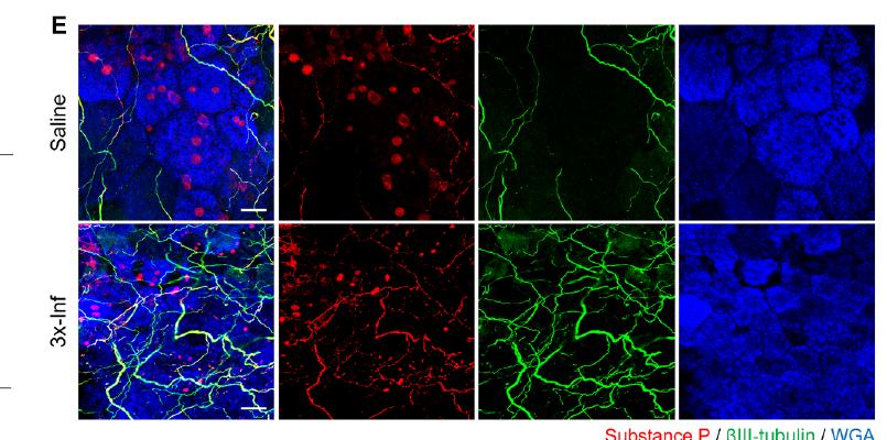
Fig.1E:whole-mount bladder imaging 显示感染后 nociceptive fibers 更密集地靠近/进入 urothelium。
Fig.1A-C(原文):公开导读版从略;完整图见 Cell 原文。
读图要点:rUPEC protocol、von Frey pain response 和 void spot dysfunction 建立慢性疼痛/排尿表型入口。
Fig.1F-I(原文):公开导读版从略;完整图见 Cell 原文。
读图要点:DEG/GO/GSEA 把长期 remodeling 指向 inflammatory cytokine 与 IL-6-JAK-STAT3 signaling。
Fig.2 | 感染后的 BEC 重塑伴随 PIEZO1-dependent Ca2+ mechanosensation
mechanosensation上皮结构/屏障改变后,PIEZO1 是感染诱导最明显的机械/Ca2+ channel;PIEZO1-deficient BEC 对 stretch 的 Ca2+ influx 明显下降。
Fig.2A-C(原文):公开导读版从略;完整图见 Cell 原文。
读图要点:mucosa separation、ZO-1/CK20/WGA 和 TEER 说明感染后 barrier/differentiation remodeling。
Fig.2D-F(原文):公开导读版从略;完整图见 Cell 原文。
读图要点:ex vivo Ca2+ signals 与 PIEZO1 expression 将上皮重塑接到 mechanosensation。
Fig.2G-H(原文):公开导读版从略;完整图见 Cell 原文。
读图要点:PIEZO1 CRISPR KO + stretch chamber 证明 Ca2+ response 的 PIEZO1 dependency。
Fig.3 | IL-6 通过 STAT3 上调 PIEZO1,并放大机械刺激 Ca2+ 反应
IL-6 primingIL-6 在感染 BEC/膀胱中升高,rhIL-6 dose-dependently 上调 PIEZO1;STAT3 motif deletion 降低 promoter response,IL-6 preconditioning 放大 YODA1/stretch Ca2+ influx。
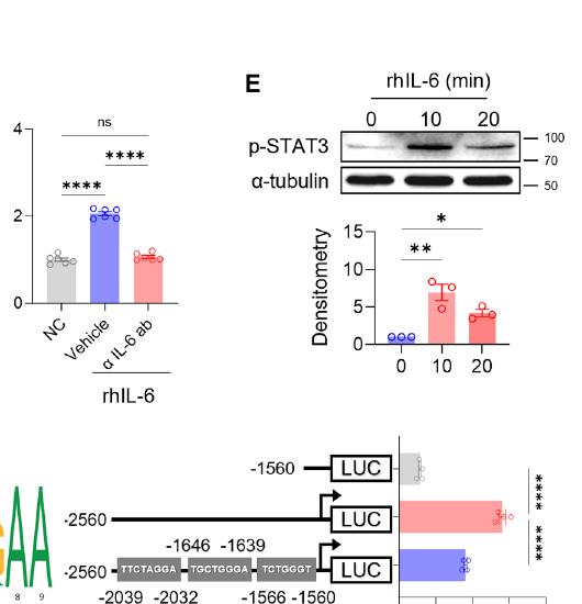
Fig.3E-F:pSTAT3 与 PIEZO1 promoter luciferase 证明 IL-6/STAT3 transcriptional priming。
Fig.3A-D(原文):公开导读版从略;完整图见 Cell 原文。
读图要点:IL-6 induction、PIEZO1 qPCR/blot 和 anti-IL-6 blockade 支撑炎症 priming。
Fig.3G-I(原文):公开导读版从略;完整图见 Cell 原文。
读图要点:YODA1/stretch/ex vivo Ca2+ 功能 panel 证明 IL-6 preconditioning 放大机械敏感性。
Fig.4 | PIEZO1-mediated Ca2+ influx 引发 ROS,并激活 NRF2 antioxidant program
redoxYODA1/stretch 增加 ROS,PIEZO1 KO 消除该反应;NRF2 nuclear translocation 和 injury scRNA/bulk deconvolution 说明 cycling/superficial urothelial cells 启动 antioxidant program。
Fig.4A-C(原文):公开导读版从略;完整图见 Cell 原文。
读图要点:YODA1/stretch 的 PIEZO1-dependent ROS production 连接 Ca2+ 与 redox stress。
Fig.4D-E(原文):公开导读版从略;完整图见 Cell 原文。
读图要点:NRF2 accumulation/nuclear translocation 说明 antioxidant program 被启动。
Fig.4F-H(原文):公开导读版从略;完整图见 Cell 原文。
读图要点:injury scRNA-seq 与 infected bladder deconvolution 定位 urothelial NRF2 program。
Fig.5 | NRF2-SLC7A11 抗氧化响应反而释放 glutamate,驱动 DRG sprouting
glutamateYODA1+IL-6 上调 SLC7A11;Piezo1 cKO 或 Il6 deficiency 阻断 in vivo SLC7A11 induction;SLC7A11/PIEZO1 影响 glutamate release,glutamate 通过 mGluR5 增加 eDRG neurite branching 和 capsaicin response。
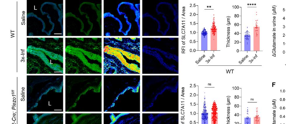
Fig.5D-F:PIEZO1-dependent in vivo SLC7A11 induction 与 urine/cell glutamate release。
Fig.5A-C(原文):公开导读版从略;完整图见 Cell 原文。
读图要点:YODA1+IL-6 induction of SLC7A11,并在 PIEZO1-deficient BEC 中消失。
Fig.5G-I(原文):公开导读版从略;完整图见 Cell 原文。
读图要点:glutamate/mGluR5/eDRG branching 和 capsaicin response 建立神经重塑下游。
Fig.6 | PIEZO1-SLC7A11 轴决定 C-fiber rewiring 和 pelvic pain
pain感染上调 Calca/CGRP 等 nociceptive markers;Slc7a11 cKO、Piezo1 cKO 或 Il6-/- 均削弱 epithelial sensory sprouting 和 von Frey hypersensitivity。
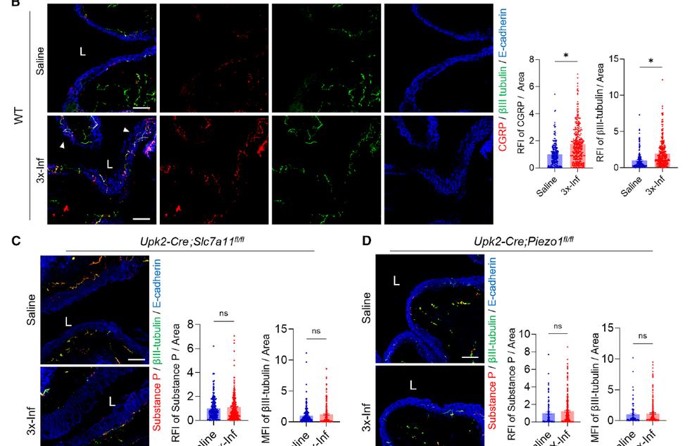
Fig.6B-D:WT 中 CGRP+/substance P+ sensory sprouting,被 Slc7a11/Piezo1 epithelial cKO 阻断。
Fig.6A(原文):公开导读版从略;完整图见 Cell 原文。
读图要点:nociceptive marker qPCR 区分 C-fiber-associated rewiring。
Fig.6E-H(原文):公开导读版从略;完整图见 Cell 原文。
读图要点:von Frey pain behavior 在 Slc7a11/Piezo1 epithelial cKO 后降低。
Fig.7 | PIEZO1-SLC7A11 轴还控制 voiding dysfunction,SSZ 可作为治疗入口
therapyPiezo1/Slc7a11 epithelial cKO 减少感染后 voiding spot/cystometry 异常;SSZ 抑制 glutamate release,并改善 von Frey pain response 和 void spot phenotype。
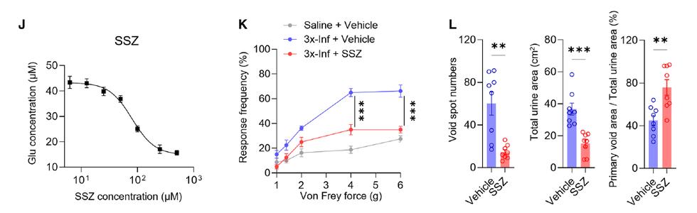
Fig.7J-L:SSZ inhibits glutamate release and rescues pain/voiding readouts。
Fig.7A(原文):公开导读版从略;完整图见 Cell 原文。
读图要点:Piezo1/Slc7a11 epithelial cKO 减少 infected voiding spot abnormalities。
Fig.7B-I(原文):公开导读版从略;完整图见 Cell 原文。
读图要点:awake cystometry 解析 contraction interval、pressure 与 void volume。
Supplementary Figures:代表图与证据索引
公开版仅保留 S12A 这个低分辨率 panel-group 作为治疗/药理补充代表图;S1-S11 以证据索引形式说明它们分别支撑哪一步,不重发布全部补图。
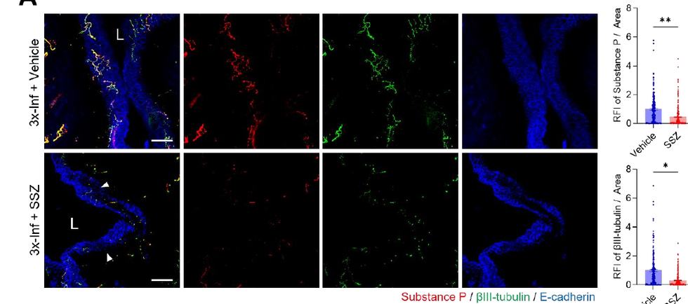
Figure S12A:SSZ 对感染后 nerve sprouting 的补充 rescue 证据。
Figure S1-S11(原文):公开导读版从略;完整图见 Cell 原文。
读图要点:S1-S11 覆盖 strain generalization、persistent neuroplasticity、uroplakin/TEER/Ca2+ validation、genetic validation、mGluR5/eDRG、cystometry/EUS-EMG 等控制实验。
Figure S1(原文):公开导读版从略;完整图见 Cell 原文。
读图要点:NU14/UTI89/CI5 strain generalization、void spot 和 TH sympathetic distribution control。
Figure S2(原文):公开导读版从略;完整图见 Cell 原文。
读图要点:2-week persistence、RNA-seq quality/scatter/MA/GSEA,支撑 Fig.1 的长期 remodeling。
Figure S3(原文):公开导读版从略;完整图见 Cell 原文。
读图要点:uroplakin reduction,补充 Fig.2 的 terminal differentiation/barrier phenotype。
Figure S4(原文):公开导读版从略;完整图见 Cell 原文。
读图要点:TEER setup 和 YODA1 Ca2+ imaging validation。
Figure S5(原文):公开导读版从略;完整图见 Cell 原文。
读图要点:inflammatory cytokines、ROS positive control 和 scRNA deconvolution controls。
Figure S6(原文):公开导读版从略;完整图见 Cell 原文。
读图要点:Piezo1 cKO、Il6-/- SLC7A11 和 SLC7A11 CRISPR validation。
Figure S7(原文):公开导读版从略;完整图见 Cell 原文。
读图要点:L6-S1 DRG mGluR5 expression 和 shRNA knockdown validation。
Figure S8(原文):公开导读版从略;完整图见 Cell 原文。
读图要点:ATP release/control、glutamate vs ATP eDRG branching、capsaicin response validation。
Figure S9(原文):公开导读版从略;完整图见 Cell 原文。
读图要点:Slc7a11 genetic validation 和 Il6-/- pain/neuroplasticity controls。
Figure S10(原文):公开导读版从略;完整图见 Cell 原文。
读图要点:Slc7a11/Piezo1 cKO voiding spot 和 awake cystometry setup。
Figure S11(原文):公开导读版从略;完整图见 Cell 原文。
读图要点:EUS-EMG 与 bladder pressure coupling,验证 detrusor-sphincter dyssynergia。
材料完整性、补充材料与数据索引
Phase 0 preflight 已按 pipeline 运行,并在人工复核后解除 blocker:主文 PDF 可读,`mmc2.pdf` 是主文重复件,`mmc1 (1).pdf` 是 24 页补充材料。
2 个主文重复件 + 1 个补充 PDF。
没有 XLSX source-data 包。
S1-S12 支撑主文图的 controls / validation。
PDF role classification
- Main paper PDF
58 pages, unencrypted; contains the Cell corrected-proof article plus repeated supplemental/STAR Methods pages.
- mmc2.pdf
Byte-identical duplicate of the main paper PDF; same SHA-256 as the long titled PDF, excluded as duplicate.
- mmc1 (1).pdf
24-page supplemental information PDF; contains Figure S1-S12, Table S1-S3 and supplemental references.
Supplementary tables
- Table S1
sgRNA oligonucleotides for PIEZO1 and SLC7A11 CRISPR targeting.
- Table S2
shRNA oligonucleotides for scramble and mGluR5 knockdown.
- Table S3
qRT-PCR primers for human and mouse targets, including PIEZO1, TRPV4, IL-1beta, TNFalpha, IL-6, SLC7A11 and antioxidant/pain markers.
Preflight manual-review notes
- Preflight verdict
Official preflight initially returned NEEDS REVIEW because all three PDFs were heuristically classified as main-paper candidates.
- Manual resolution
The long titled PDF and mmc2.pdf are exact duplicates; mmc1 is the standalone supplement. No encrypted or unreadable core file was found.
- No source workbooks
This folder has no XLSX source-data workbook; the source-data section is therefore a PDF/table inventory, not a workbook sheet map.
总结与延伸
SLC7A11 的 antioxidant homeostasis 在上皮内部是保护性的,但它的 glutamate efflux 在组织层面变成 sensory remodeling signal。
文章真正研究的是 urothelium-neuron interface,而不是单独的 epithelial inflammation 或单独的 pain neuron excitability。
每个核心节点都有上游/下游 perturbation:IL-6 antibody、STAT3 promoter deletion、PIEZO1 KO/cKO、SLC7A11 KO/cKO、mGluR5 shRNA、SSZ/SORA。
SSZ/SORA 不只是镇痛,而是试图从上皮代谢输出处减少 neuroplasticity driver,这比单纯抑制神经受体更 upstream。
最值得复用的实验结构
- 把临床症状拆成结构、分子、神经和器官功能四个层级,并在每层设遗传 rescue。
- 同一条轴使用体外 BEC、ex vivo mucosal sheet、in vivo cKO 和 primary/eDRG neuron 多系统交叉验证。
- 将 RNA-seq/GSEA/scRNA reference deconvolution 用作定位假说,而不是让组学替代功能实验。
- 治疗实验不仅看 pain response,还看 nerve sprouting、glutamate release、void spot 和 cystometry。
不足、风险与开放问题
- 模型边界:反复腔内 UPEC instillation 不能完全模拟临床 antibiotic clearance 后 recurrence,但适合研究 repeated epithelial stimulation。
- 时间定义:文章强调 sustained pain-like state 和 neuroepithelial remodeling;不能直接等同所有人类慢性盆腔痛诊断标准。
- IL-6 干预风险:IL-6 是 upstream priming,但感染背景下全身阻断可能影响 host defense;SLC7A11/glutamate output 可能更适合作为局部/下游入口。
- glutamate 之外:NGF、BDNF、ATP、immune mediators 可能并行参与 sensory plasticity;本文把 glutamate 证明为 dominant structural remodeling driver,但不排除协同网络。
- 下一步:可用 human rUTI/IC biospecimens 做 urothelial PIEZO1/SLC7A11/glutamate/mGluR5 spatial validation,并做 longitudinal symptom correlation。